


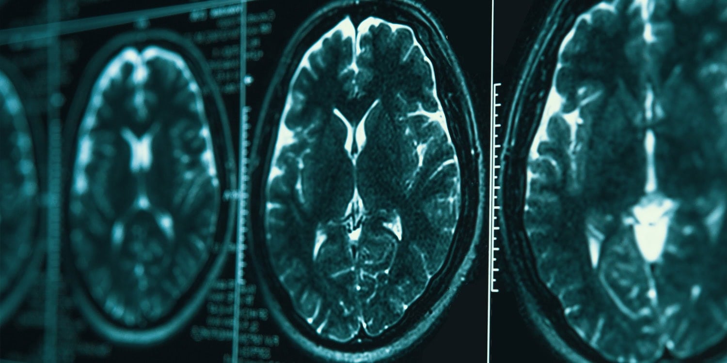
A new, small exploratory study links the structure of the brain’s outer layer to how well patients with major depressive disorder respond to the antidepressant vortioxetine, measured before treatment began and assessed after around eight weeks. The research identifies several regions implicated in emotion regulation, visual processing, and bodily awareness as potential predictors of symptom improvement.
Major depressive disorder is a common condition that can markedly disrupt daily life, and remission after initial treatment remains elusive for many patients. The study adds to efforts to personalise care by seeking brain-based indicators of which treatments might work best for an individual.
Researchers at Beijing HuiLongGuan Hospital in China used high-resolution structural magnetic resonance imaging, focusing on the brain’s anatomy rather than activity, to examine whether features of the cortex could forecast improvement in depressive symptoms after antidepressant therapy. They also explored whether cognitive changes accompany mood improvements, with a particular interest in memory.
The team recruited 34 adults aged 18 to 50 with major depressive disorder and 26 healthy controls matched for age and sex. All patients had experienced prior depressive episodes and had not taken antidepressants in the eight weeks before joining the study. After accounting for data quality and follow-up attendance, analyses were conducted on 22 patients and 21 controls.
Participants with depression were given a daily 10-milligram dose of vortioxetine. They underwent assessments before and after treatment, including the 17-item Hamilton Rating Scale for Depression and the Hamilton Anxiety Rating Scale, as well as the Repeatable Battery for the Assessment of Neuropsychological Status to test memory, attention, language, and visual-spatial skills. On average, depressive symptoms fell from about 27 to around 8 over the treatment period.
All patient participants had brain scans at the start and again about eight weeks later; the healthy controls were scanned once. The analysis, however, utilised only the baseline scans to see whether pretreatment brain features were linked to subsequent improvement.
The researchers examined four cortical surface features: thickness; the gyrification index, reflecting how folded the cortex is; sulcus depth, the depth of grooves between folds; and fractal dimension, a measure of the complexity of folding patterns.
Compared with healthy participants, those with depression showed less folding in the right insula, a region involved in emotional awareness and body state monitoring.
Among participants with depression, higher fractal dimension—more complex cortical folding—in the left superior frontal cortex, the right rostral middle frontal cortex, and the right lateral occipital cortex was associated with greater symptom improvement. The frontal areas are part of the prefrontal cortex, linked to emotion regulation and executive control, while the lateral occipital cortex is a visual region. Greater cortical thickness in the insula on both sides and in the left pericalcarine cortex, part of the primary visual cortex, was also linked to greater improvement.
Conversely, lower fractal dimension in the left cuneus, another visual region, and less folding in the left lateral occipital cortex were associated with greater improvement. Several of these associations, including those involving the insula and the right lateral occipital cortex, related to improvements in anxiety scores rather than depressive symptoms.
On the cognitive side, shallower grooves in the left lateral occipital cortex (lower sulcus depth) were linked to greater improvement in delayed memory, the ability to recall information after a delay.
To test the robustness of these findings, the researchers applied leave-one-out cross-validation, building models on all but one participant and then predicting the remaining case. In these analyses, fractal dimension of the left cuneus correlated with changes in depression scores (borderline statistical significance), and fractal dimension of the left superior frontal cortex correlated with changes in anxiety scores. The association between sulcus depth in the left lateral occipital cortex and delayed memory did not reach significance in this test, however.
“Baseline cortical morphometric features were associated with clinical and cognitive changes following antidepressant treatment in MDD,” the study authors stated, adding that “these findings are preliminary and should be interpreted with caution, pending replication in independent cohorts.”
The study contributes to understanding how the brain’s structure might relate to the course of depression during treatment, but the authors caution that the small sample size and multiple analyses raise the possibility of false positives. They note that results may differ in larger studies, and that the findings may not generalise to other antidepressants or to older or younger populations. There was no placebo group, so the study cannot separate the drug’s effect from other factors, and causal conclusions cannot be drawn from the design.
The paper, titled “Baseline cortical structural features are associated with depressive and cognitive improvement in major depressive disorder,” was authored by Yingna Li, Shuping Tian, Siyan Zan, Ruonan Du, Wenzhou Liang, Liying Zhao, and Zhiren Wang.
