


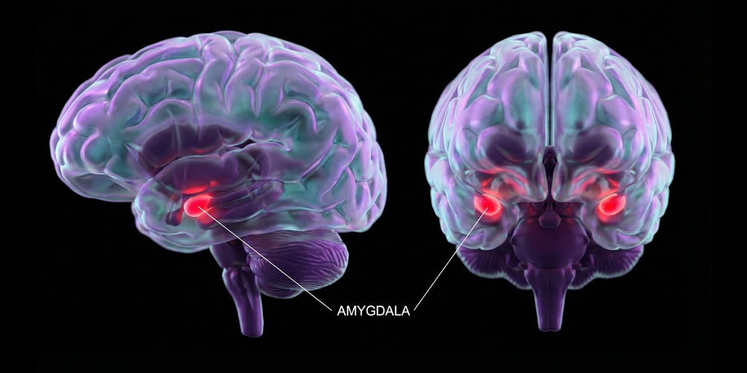
A rare clinical case published in Nature Communications reports that selectively inactivating part of the right amygdala in a man with drug‑resistant epilepsy and severe post‑traumatic stress disorder (PTSD) reduced his tendency to interpret ambiguous faces as fearful and was followed by a sharp drop in trauma symptoms. The study tracked brain signals and emotion perception before and immediately after an awake bedside procedure, offering unusual causal evidence that right‑amygdala activity helps sustain threat‑focused biases and PTSD symptoms.
Understanding threat bias and the amygdala
PTSD can develop after severe trauma and is characterised by intrusive memories, avoidance of reminders, negative shifts in mood and heightened physical arousal. A central feature is a negative interpretation bias—the tendency to read neutral or ambiguous cues, such as an unreadable facial expression, as threatening. This bias can keep fear responses active and complicate emotional regulation.
The amygdala—a pair of almond‑shaped clusters deep in the brain’s temporal lobes—has long been the focus of research into threat processing. Brain scans often show heightened amygdala reactivity to fearful or neutral cues in people with PTSD, but standard imaging only reveals correlations. The question has been whether abnormal amygdala activity drives negative interpretation bias or merely accompanies it.
Studying cases of localized brain damage or targeted neurosurgery provides a rare way to observe what happens when amygdala function is directly disrupted. In people without PTSD, bilateral amygdala damage can blunt recognition of fearful faces while leaving happy-face recognition intact. Clinically, patients treated for limbic epilepsy—where seizures arise in deep emotion and memory structures such as the amygdala and hippocampus—offer a roadmap for linking psychiatric symptoms to specific brain circuits.
Mapping emotion and ablating the right amygdala
The patient was a 41‑year‑old man with drug‑resistant temporal lobe epilepsy and severe PTSD from adolescent trauma. To determine the amygdala’s role, doctors used stereoelectroencephalography‑guided radiofrequency ablation, implanting thin wire electrodes to locate seizure sources, then inactivating selected tissue with heat while the patient remained awake.
Before the ablation, the team compared him with 16 temporal lobe epilepsy patients undergoing similar monitoring but without PTSD. They completed 252 trials of a facial emotion discrimination task, where faces morphed from fully happy to fully fearful and participants judged each face while rating confidence. Recordings captured late‑phase brain activity 350 to 650 milliseconds after each face, reflecting sustained emotional processing.
On the morning of the procedure, researchers mapped the patient’s amygdala with brief electrical pulses, asking him to report sensations. That afternoon, doctors ablated tissue around five contacts in his right basal and lateral amygdala, and 20 minutes later he completed 336 trials of the face task.
Post‑ablation MRI showed that the lesion inactivated 47 percent of the right amygdala while sparing more than 99.9 percent of the right hippocampus. Electrophysiological data confirmed the targeted tissue ceased producing normal activity during recording sessions.
After the ablation, the patient’s responses shifted. He judged ambiguous 50‑percent fearful faces as fearful much less often, aligning with control participants, and his reactions to moderately happy faces showed no significant change. His overall speed of decision‑making increased more slowly, and his confidence patterns during the task also shifted, erasing the pre‑ablation bias toward judging fearful faces as more probable.
Notably, the late‑phase gap between fearful and happy face responses vanished across the right amygdala and nearby right‑hemisphere areas. A new pattern emerged in his unaffected left amygdala, alongside smaller responses to ambiguous faces, suggesting that silencing threat signals from the right amygdala may redirect emotion processing toward the left amygdala and slower, more deliberate evaluation.
Two months after the procedure, validated questionnaires showed a dramatic improvement in PTSD symptoms, with the PTSD score dropping from 50 (severe) to 21, well below the diagnostic threshold. Intrusive memories, avoidance, negative mood, and hyperarousal all improved, with milder reductions in anxiety and depression. Seizure frequency remained around 10 per month during that period, rising to fewer than one per month only after a second, more extensive laser surgery on the right hippocampus and entorhinal region.
Crucially, the PTSD relief occurred independently of seizure control, and the patient was not undergoing structured talk therapy or PTSD‑targeted medications during the study. “The remarkable result was evidence of immediate widespread changes in behavior, brain dynamics, and PTSD benefit without dependency on other traditional therapies such as medications or cognitive behavioural therapy,” Willie explained. “This is an early feasibility study that could lead to future larger studies that target the right amygdala to treat PTSD.”
The findings align with earlier work in brain circuits and neurostimulation among veterans, which suggested that dampening amygdala‑linked pathways can ease PTSD symptoms. They also echo a prior functional MRI study involving identical twins that linked heightened amygdala reactivity to threat processing of ambiguous expressions, though that work was correlational and not based on surgical intervention in a single patient.
Senior study author Jon T. Willie, chief of functional and epilepsy surgery at the University of Texas at Austin’s Dell Medical School, emphasised the novelty of the work. “The novelty of the study is that this is the first clear demonstration of a specific causal role for the right amygdala in maintaining PTSD, indicating that therapies that reduce or block right amygdala could provide effective therapy for PTSD,” he said. He also cautioned that the study remains early and suggests the need for larger trials to determine safety and feasibility across patient groups.
There are important caveats. The evidence comes from a single patient whose brain circuitry was shaped by years of epilepsy and antiseizure medications, so his neural organisation may differ from most people with PTSD. The researchers note that amygdala ablation has not been widely studied as a therapy for PTSD, and awake surgery cannot include a blinded sham procedure, which could influence performance. The team acknowledges that ablating right amygdala tissue may blunt overall emotional calibration rather than solely removing threat bias.
Because the patient underwent subsequent right‑hippocampus surgery two months later, the durability of the right‑amygdala intervention beyond that period remains unclear. Nevertheless, PTSD relief persisted at least a year after the initial procedure. The researchers advocate larger prospective trials and the exploration of reversible techniques such as focused ultrasound or targeted electrical stimulation to test whether modulating the right amygdala can safely treat severe PTSD.
Looking ahead, the team is studying how the amygdala shapes the brain’s ability to recognise safety versus threat, with plans to test mechanisms of fear and safety learning in epilepsy patients undergoing intracranial monitoring. “Our next steps are to better define the mechanisms by which amygdala‑focused interventions can be used to understand and treat PTSD,” Willie added.
The study’s authors are Tao Xie, Sanne J. H. van Rooij, Sai Sun, Noah K. Bryson, Phillip Demarest, Haeorum Park, Luigi Maccotta, Shuo Wang, Peter Brunner, and Jon T. Willie.
