


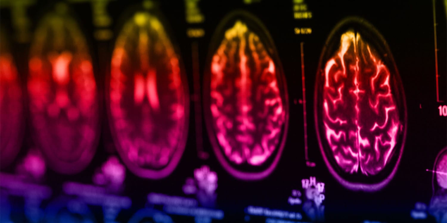
A new analysis of brain imaging data suggests that ADHD may influence brain iron levels in relation to when children start stimulant medication. The study found that starting treatment by age eight is linked with higher iron concentrations in dopamine-rich brain regions than starting at age nine or later, and the findings are published in Brain Research.
Attention deficit hyperactivity disorder is a neurodevelopmental condition characterised by inattention, impulsivity and hyperactivity. These symptoms often complicate daily tasks and social interactions, and the condition is closely tied to reduced dopamine signaling in the brain. Psychostimulants, commonly prescribed to manage ADHD, work by increasing the amount of dopamine available in the central nervous system.
Brain iron plays an essential role in dopamine production, as tyrosine hydroxylase—the enzyme responsible for creating dopamine—relies on iron to function. Iron also supports myelin production, which speeds up communication between brain cells. Consequently, iron tends to concentrate in dopamine-rich regions such as the basal ganglia and the substantia nigra, deep structures integral to motor control and learning. Iron levels in these areas typically rise as children mature and the dopamine system develops.
Previous work has suggested that untreated ADHD may be associated with lower brain iron compared with neurotypical peers, while some imaging studies have reported iron levels in medicated children may resemble those of non-affected individuals. This raised questions about whether stimulant medications may modify brain iron levels over time.
To investigate, Hugo A.E. Morandini of the Child and Adolescent Health Service in Perth, Australia, and colleagues designed a two-year longitudinal study examining how the duration of treatment and the age at which treatment began related to brain iron. The team analysed brain imaging data from the Oregon ADHD-1000 dataset, a public clinical repository, identifying 29 children with ADHD who were consistently taking psychostimulants and 38 neurotypical children with no psychiatric diagnoses. Scans were conducted at the outset and repeated two years later.
The researchers employed functional magnetic resonance imaging and a specialised analysis to estimate brain iron concentration based on tissue magnetic properties. They focused on several regions rich in both dopamine and iron, including the globus pallidus, putamen, caudate nucleus, thalamus and substantia nigra.
Across the two-year period, the study found no statistically significant differences in iron levels between children with ADHD and neurotypical peers in the substantia nigra, thalamus and most of the basal ganglia. However, those with ADHD showed lower iron content in the globus pallidus, a key part of the basal ganglia involved in regulating dopamine circuitry and reward processing. The authors suggested that the shorter average duration of medication exposure in their sample compared with earlier work could help explain why globus pallidus iron had not equalled that of neurotypical peers.
Within the ADHD group, the researchers assessed how treatment history related to brain iron. They calculated the total time each child had been on stimulants and noted the age at which treatment started, then tested whether these factors predicted iron levels in the different regions. The duration of stimulant use showed little relation to brain iron, with only a very small negative association seen in the thalamus that the team judged clinically unlikely to matter.
By contrast, the age at which treatment began produced a clearer pattern. The ADHD children were divided into two groups: those who started eight or younger and those who started at nine or older. The earlier-start group exhibited higher iron content in the thalamus, caudate nucleus, putamen and globus pallidus. The authors proposed that early childhood is a period of rapid developmental changes in iron and dopamine, and that starting medication earlier may intersect with these windows, potentially increasing the brain’s sensitivity to pharmacological influences.
The study does acknowledge several limitations. The sample size was modest, and a larger study would help clarify the strength and precision of these associations. Treatment timelines were based on parental reports, which can be imprecise, and there was no detailed information on medication dosages or adherence. Because the research observed existing prescribing patterns rather than assigning treatments, the findings indicate association rather than causation, and they cannot fully disentangle underlying symptom severity from medication effects. Measuring brain iron before treatment would also be needed to isolate drug-specific impacts.
Looking ahead, the researchers call for longer-term studies that follow patients from before they begin treatment into adulthood to map how early stimulant use may shape brain iron over the lifespan and whether observed differences persist into later life. The study, Neurochemical impact of psychostimulants: Longitudinal MRI analysis of brain iron content in children with ADHD, was authored by Hugo A.E. Morandini, Sjoerd B. Vos, Paola Chivers, Tim J. Silk and Pradeep Rao.
